Help ?

IGMIN: We're glad you're here. Please click 'create a new query' if you are a new visitor to our website and need further information from us.

If you are already a member of our network and need to keep track of any developments regarding a question you have already submitted, click 'take me to my Query.'

Search

Organised by  IgMin Fevicon

Regional sites

Browse by Subjects

Welcome to IgMin Research – an Open Access journal uniting Biology, Medicine, and Engineering. We’re dedicated to advancing global knowledge and fostering collaboration across scientific fields.

Browse by Sections

At IgMin Research, we bridge the frontiers of Biology, Medicine, and Engineering to foster interdisciplinary innovation. Our expanded scope now embraces a wide spectrum of scientific disciplines, empowering global researchers to explore, contribute, and collaborate through open access.

Members

Our purpose is to inspire cross-field connections that drive new insights and developments.

Articles

Our purpose is to inspire cross-field connections that drive new insights and developments.

Explore Content

Our purpose is to inspire cross-field connections that drive new insights and developments.

Identify Us

Our purpose is to inspire cross-field connections that drive new insights and developments.

IgMin Corporation

Welcome to IgMin, a leading platform dedicated to enhancing knowledge dissemination and professional growth across multiple fields of science, technology, and the humanities. We believe in the power of open access, collaboration, and innovation. Our goal is to provide individuals and organizations with the tools they need to succeed in the global knowledge economy.

Publications Support
[email protected]
E-Books Support
[email protected]
Webinars & Conferences Support
[email protected]
Content Writing Support
[email protected]
IT Support
[email protected]

Search

Select Language

Explore Section

Content for the explore section slider goes here.

Abstract

Abstract at IgMin Research

Our purpose is to inspire cross-field connections that drive new insights and developments.

Engineering Group Research Article Article ID: igmin307

Melanocytic Nevi Classification using Transfer Learning

Machine Learning Artificial Intelligence DOI10.61927/igmin307 Affiliation

Affiliation

    1Associate Professor, Department of CSE (AI&ML), ATME College of Engineering, Mysore, Karnataka, India

    2Department of CSE (AI&ML), ATME College of Engineering, Mysore, Karnataka, India

58
VIEWS
23
DOWNLOADS
Connect with Us

Abstract

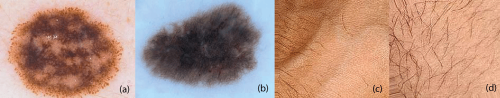
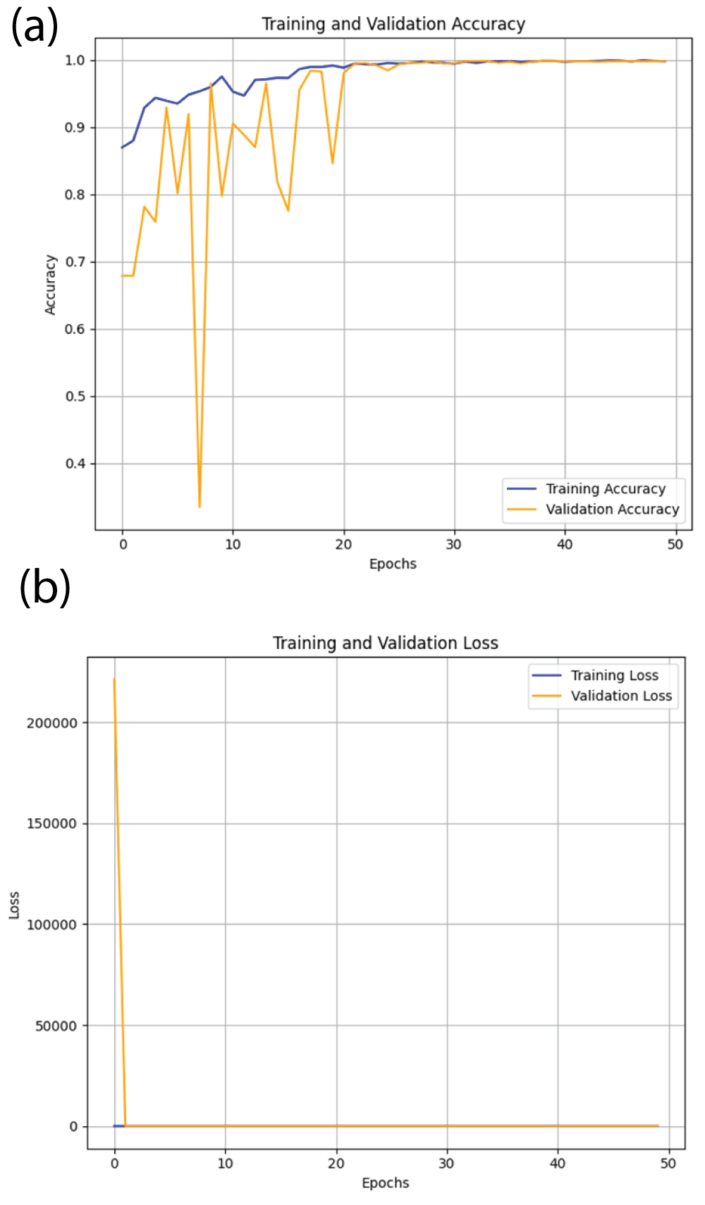
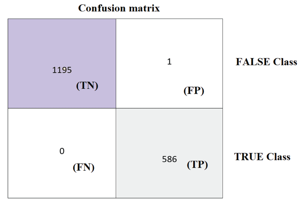
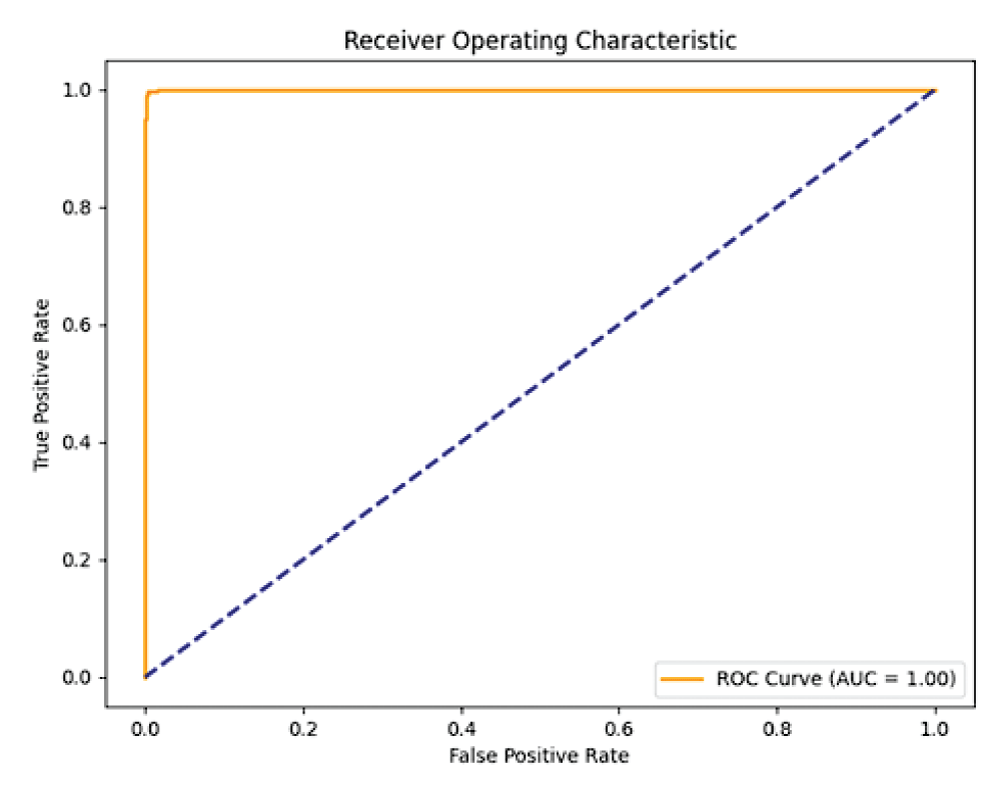
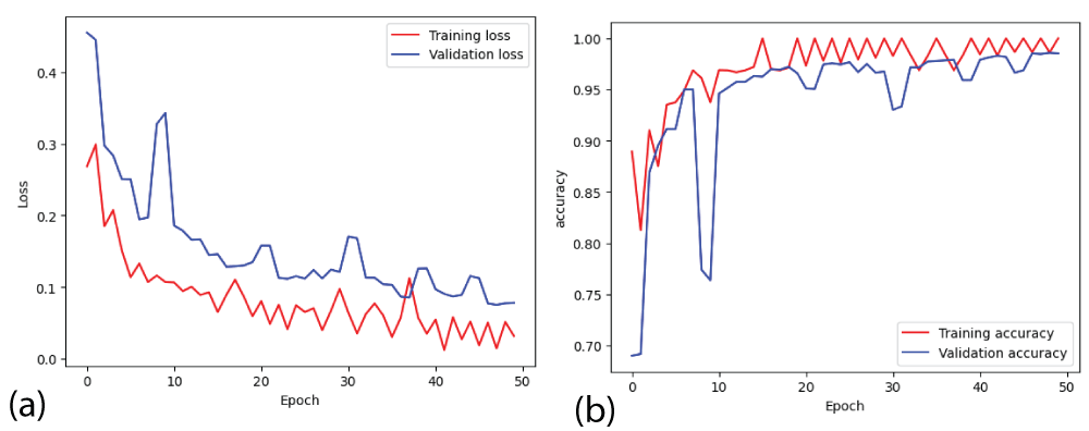
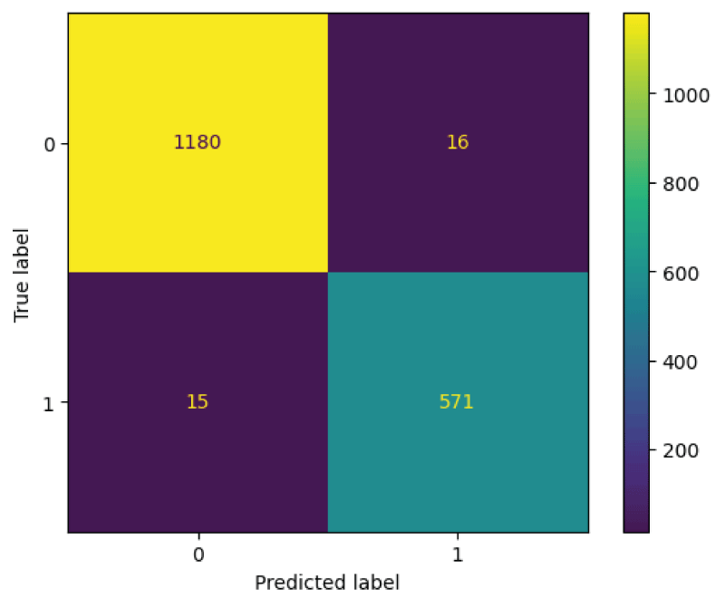
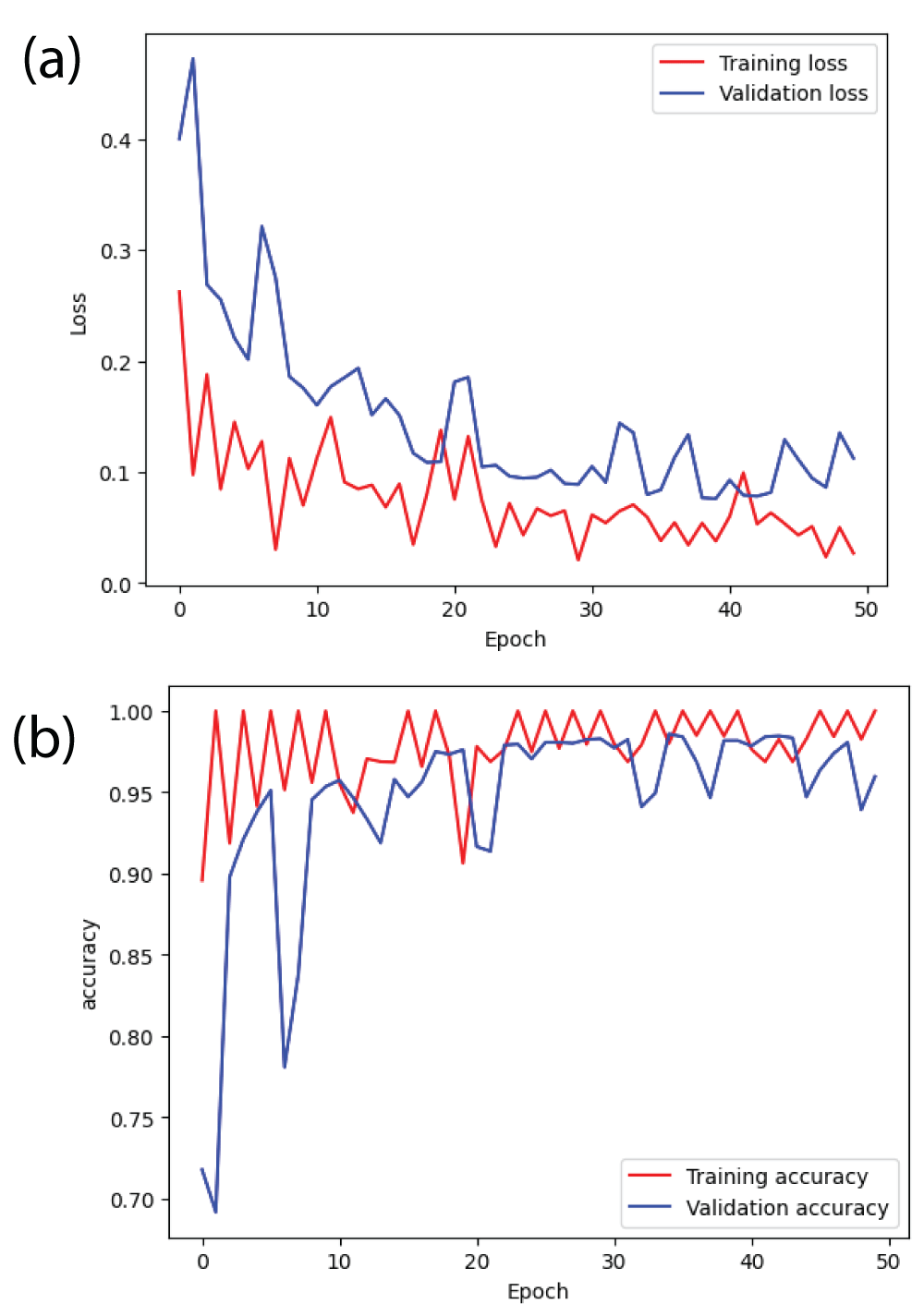
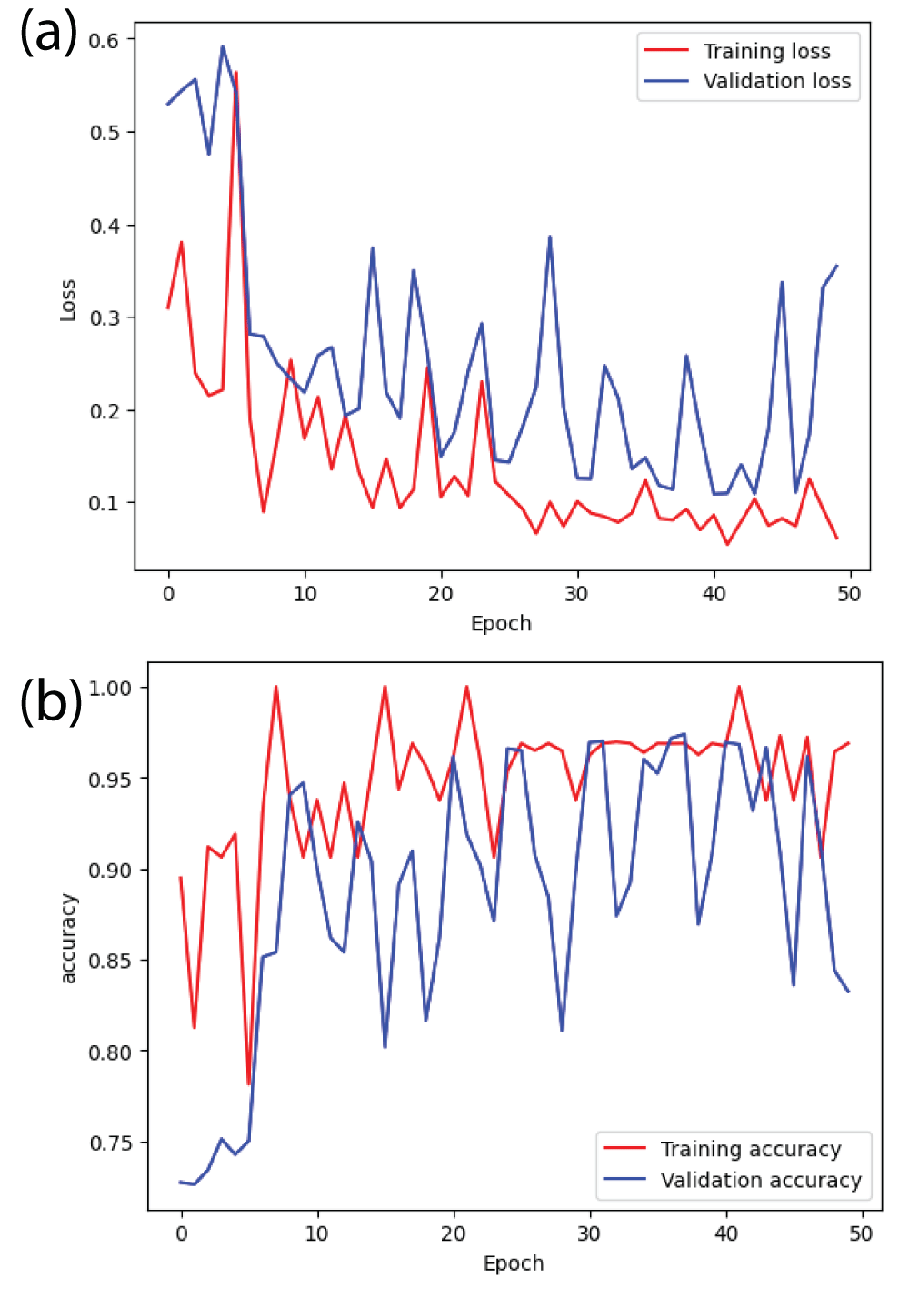
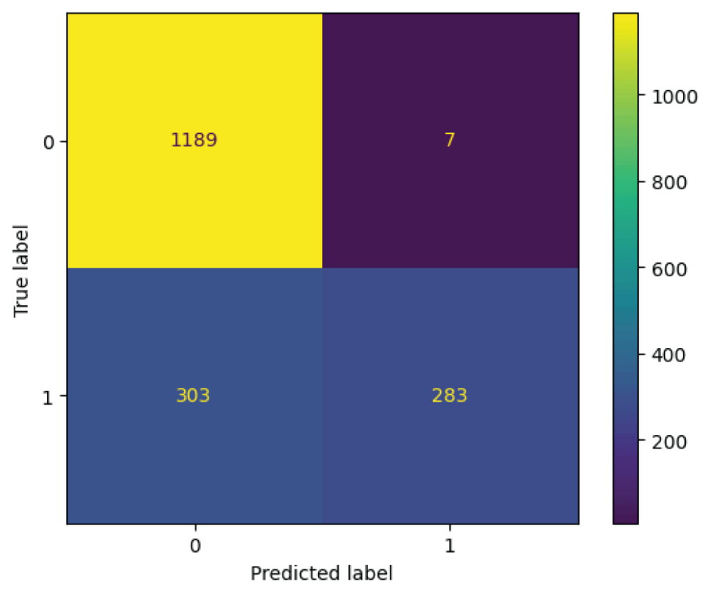
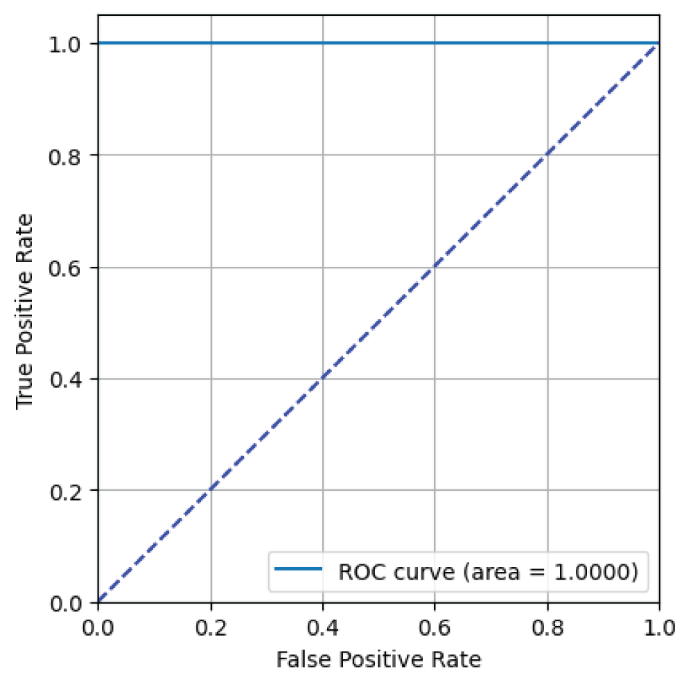
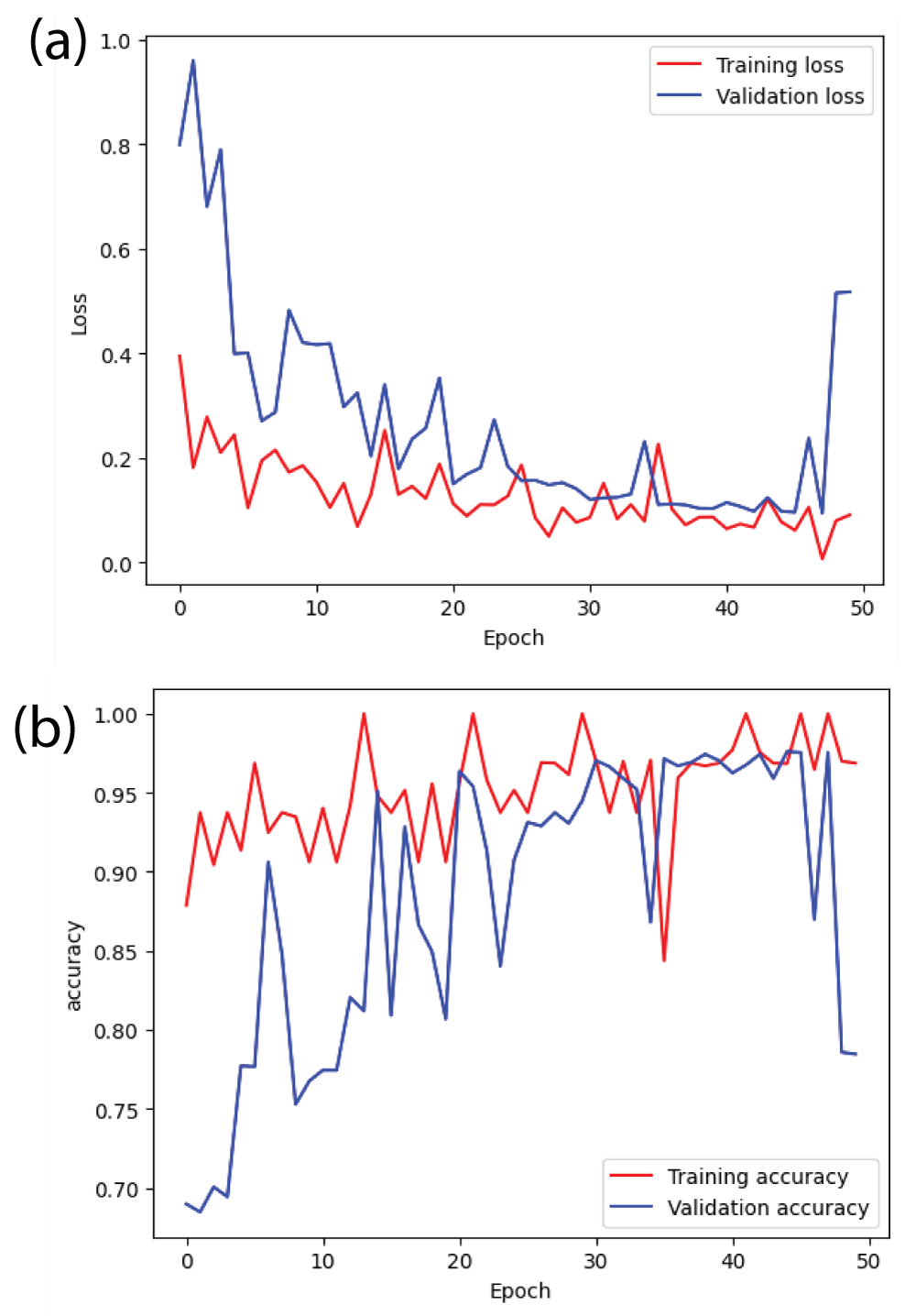
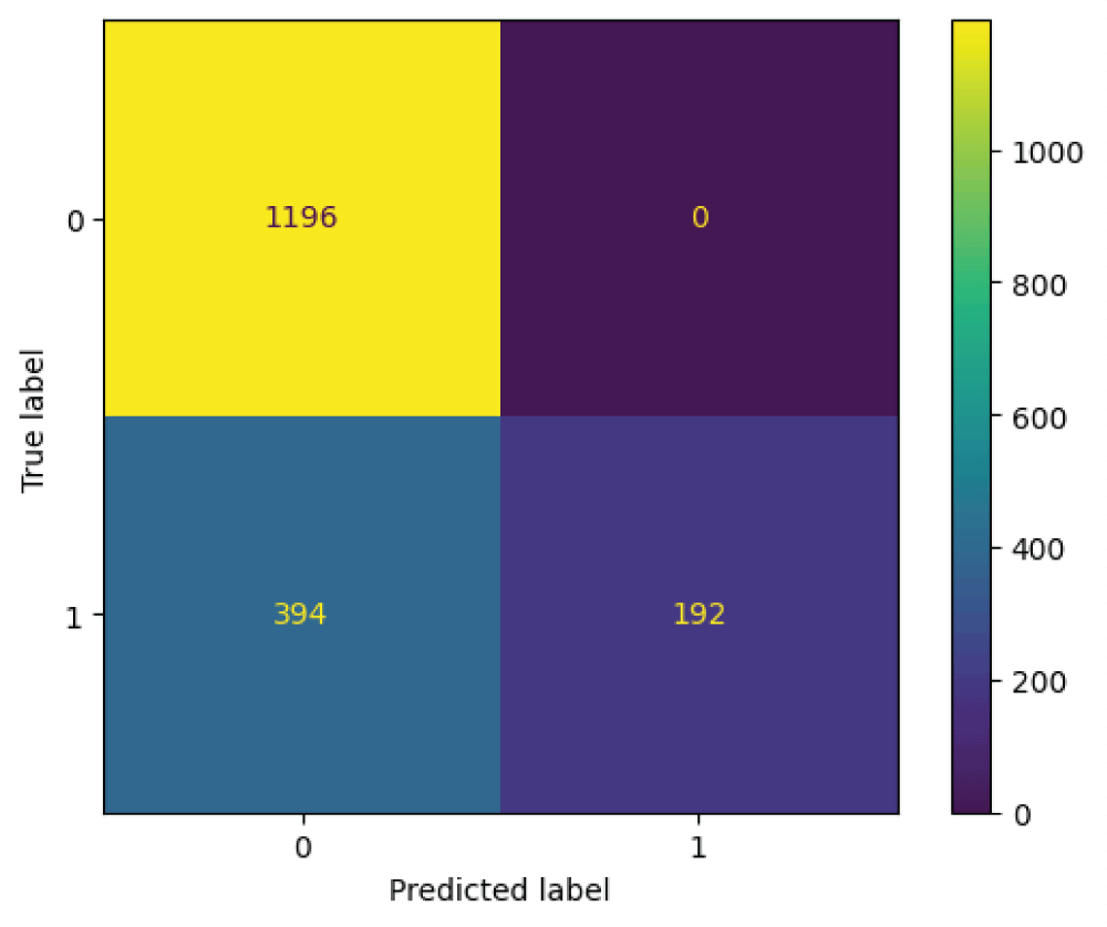
In this paper, the binary classification of skin images has been performed using deep learning technique. i.e the skin disease recognition has been performed using deep learning technique. Here, the binary classification of skin images namely melanocytic nevi and normal skin images has been classified using resnet50 deep learning network. Normal skin images have been considered in TRUE class. Melanocytic nevi skin images have been considered in FALSE class. Traditional method such as biopsy involves lot of computational procedures and consumes a lot of time which is tedious process. Therefore, deep learning-based skin disease recognition has been proposed here. The dataset for melanocytic nevi and normal skin images have been prepared for 9792 images. This dataset is passed through all five deep residual network models to obtain the results. The results such as error/accuracy curves, error matrix, false-positive-rate (FPR) vs. true-positive-rate (TPR) curve are shown for the confirmation of the work. The results obtained from the ResNet50 model were compared with other deep residual network models i.e ResNet18, ResNet34, ResNet101, and ResNet152 models.

Figures

References

    1. Huang JM, Chikeka I, Hornyak TJ. Melanocytic Nevi and the Genetic and Epigenetic Control of Oncogene-Induced Senescence. Dermatol Clin. 2017 Jan;35(1):85-93. doi: 10.1016/j.det.2016.08.001. PMID: 27890240; PMCID: PMC5391772.
    2. Gandini S, Sera F, Cattaruzza MS, Pasquini P, Abeni D, Boyle P, Melchi CF. Meta-analysis of risk factors for cutaneous melanoma: I. Common and atypical naevi. Eur J Cancer. 2005 Jan;41(1):28-44. doi: 10.1016/j.ejca.2004.10.015. PMID: 15617989.
    3. Harvey NT, Wood BA. A Practical Approach to the Diagnosis of Melanocytic Lesions. Arch Pathol Lab Med. 2019 Jul;143(7):789-810. doi: 10.5858/arpa.2017-0547-RA. Epub 2018 Jul 30. PMID: 30059258.
    4. Jibhakate A, Parnerkar P, Mondal S, Bharambe V, Mantri S. Skin lesion classification using deep learning and image processing. In: 2020 3rd International Conference on Intelligent Sustainable Systems (ICISS); 2020 Dec; Palladam, India. Piscataway (NJ): IEEE; 2020;333-40.
    5. Sun Q, Tang Y, Wang S, Chen J, Xu H, Ling Y. A deep learning-based melanocytic nevi classification algorithm by leveraging physiologic-inspired knowledge and channel encoded information. IEEE Access. 2024;12.
    6. Esteva A, Kuprel B, Novoa RA, Ko J, Swetter SM, Blau HM, Thrun S. Dermatologist-level classification of skin cancer with deep neural networks. Nature. 2017 Feb 2;542(7639):115-118. doi: 10.1038/nature21056. Epub 2017 Jan 25. Erratum in: Nature. 2017 Jun 28;546(7660):686. doi: 10.1038/nature22985. PMID: 28117445; PMCID: PMC8382232.
    7. Kassem MA, Hosny KM, Damaševičius R, Eltoukhy MM. Machine Learning and Deep Learning Methods for Skin Lesion Classification and Diagnosis: A Systematic Review. Diagnostics (Basel). 2021 Jul 31;11(8):1390. doi: 10.3390/diagnostics11081390. PMID: 34441324; PMCID: PMC8391467.
    8. Li LF, Wang X, Hu WJ, Xiong NN, Du YX, Li BS. Deep learning in skin disease image recognition: A review. IEEE Access. 2020;8:208264–80.
    9. Theckedath D, Sedamkar RR. Detecting affect states using VGG16, ResNet50 and SE-ResNet50 networks. SN Comput Sci. 2020;1(2):79.
    10. Calderón C, Sanchez K, Castillo S, Arguello H. BILSK: A bilinear convolutional neural network approach for skin lesion classification. Comput Methods Programs Biomed Update. 2021;1:100036.
    11. Gessert N, Sentker T, Madesta F, Schmitz R, Kniep H, Baltruschat I, Werner R, Schlaefer A. Skin Lesion Classification Using CNNs With Patch-Based Attention and Diagnosis-Guided Loss Weighting. IEEE Trans Biomed Eng. 2020 Feb;67(2):495-503. doi: 10.1109/TBME.2019.2915839. Epub 2019 May 9. PMID: 31071016.
    12. Abbas Q, Celebi ME. DermoDeep—A classification of melanoma-nevus skin lesions using multi-feature fusion of visual features and deep neural network. Multimed Tools Appl. 2019;78(16):23559–80.
    13. Sinha S, Gupta N. Classification of melanocytic nevus images using BigTransfer (BiT): A study on a novel transfer learning-based method to classify melanocytic nevus images. In: 2022 International Conference on Computing, Communication, and Intelligent Systems (ICCCIS); 2022 Nov; Greater Noida, India. Piscataway (NJ): IEEE; 2022;708–12.
    14. Fraiwan M, Faouri E. On the Automatic Detection and Classification of Skin Cancer Using Deep Transfer Learning. Sensors (Basel). 2022 Jun 30;22(13):4963. doi: 10.3390/s22134963. PMID: 35808463; PMCID: PMC9269808.
    15. Xin C, Liu Z, Zhao K, Miao L, Ma Y, Zhu X, Zhou Q, Wang S, Li L, Yang F, Xu S, Chen H. An improved transformer network for skin cancer classification. Comput Biol Med. 2022 Oct;149:105939. doi: 10.1016/j.compbiomed.2022.105939. Epub 2022 Aug 10. PMID: 36037629.
    16. Mahesh U, Kiran B. Three-dimensional (3-D) objects classification by means of phase-only digital holographic information using Alex Network. In: 2024 International Conference on Signal Processing, Computation, Electronics, Power and Telecommunication (IConSCEPT); 2024 Jul; [location unspecified]. Piscataway (NJ): IEEE; 2024;1–5.
    17. Argenziano G, Zalaudek I, Ferrara G, Hofmann-Wellenhof R, Soyer HP. Proposal of a new classification system for melanocytic naevi. Br J Dermatol. 2007 Aug;157(2):217-27. doi: 10.1111/j.1365-2133.2007.07972.x. Epub 2007 Jun 6. PMID: 17553053.
    18. Kumar G, Bhatia PK. A detailed review of feature extraction in image processing systems. In: 2014 Fourth International Conference on Advanced Computing & Communication Technologies (ACCT); 2014 Feb; Rohtak, India. Piscataway (NJ): IEEE; 2014;5–12.
    19. Weiss K, Khoshgoftaar TM, Wang D. A survey of transfer learning. J Big Data. 2016;3:9.
    20. Tschandl P, Rosendahl C, Kittler H. The HAM10000 dataset, a large collection of multi-source dermatoscopic images of common pigmented skin lesions. Sci Data. 2018 Aug 14;5:180161. doi: 10.1038/sdata.2018.161. PMID: 30106392; PMCID: PMC6091241.
    21. Deng J, Dong W, Socher R, Li LJ, Li K, Fei-Fei L. ImageNet: A large-scale hierarchical image database. In: 2009 IEEE Conference on Computer Vision and Pattern Recognition (CVPR); 2009 Jun; Miami, FL. Piscataway (NJ): IEEE; 2009; 248–55.
    22. Reddy BL, Uma Mahesh RN, Nelleri A. Deep convolutional neural network for three-dimensional objects classification using off-axis digital Fresnel holography. J Mod Opt. 2022;69(13):705–17.
    23. Mahesh RU, Nagaraju S. Three-dimensional (3-D) objects classification by means of phase-only digital holographic information using deep learning. In: Data Science & Exploration in Artificial Intelligence: Proceedings of the First International Conference on Data Science & Exploration in Artificial Intelligence (CODE-AI 2024); 2024 Jul 3–4; Bangalore, India. Volume 1. Boca Raton (FL): CRC Press; 2025;363.
    24. He K, Zhang X, Ren S, Sun J. Deep residual learning for image recognition. In: Proceedings of the IEEE Conference on Computer Vision and Pattern Recognition (CVPR); 2016 Jun 27–30; Las Vegas, NV. Piscataway (NJ): IEEE; 2016;770–8. doi: 10.1109/CVPR.2016.90.

Why publish with us?

  • Global Visibility – Indexed in major databases

  • Fast Peer Review – Decision within 14–21 days

  • Open Access – Maximize readership and citation

  • Multidisciplinary Scope – Biology, Medicine and Engineering

  • Editorial Board Excellence – Global experts involved

  • University Library Indexing – Via OCLC

  • Permanent Archiving – CrossRef DOI

  • APC – Affordable APCs with discounts

  • Citation – High Citation Potential

Submit Your Article

Advertisement